X-RAY FACILITY
Professional, safe radiological examinations for the precise diagnosis of malocclusions and the creation of a treatment plan tailored to the patient's needs.
Our Orthodontic Practice has its own modern X-ray facility where our qualified and experienced staff safely and efficiently perform high-quality radiological examinations essential for diagnosis and treatment planning.
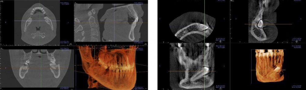
Images are visible and available immediately after they are captured. Patients can receive them on a CD for use by other specialists, e.g., for dental consultations and treatment. They can also be sent to a provided email address.
Accurate diagnostics and a precise diagnosis based upon them are prerequisites for developing an effective treatment plan.
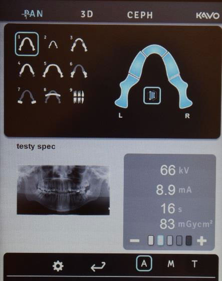
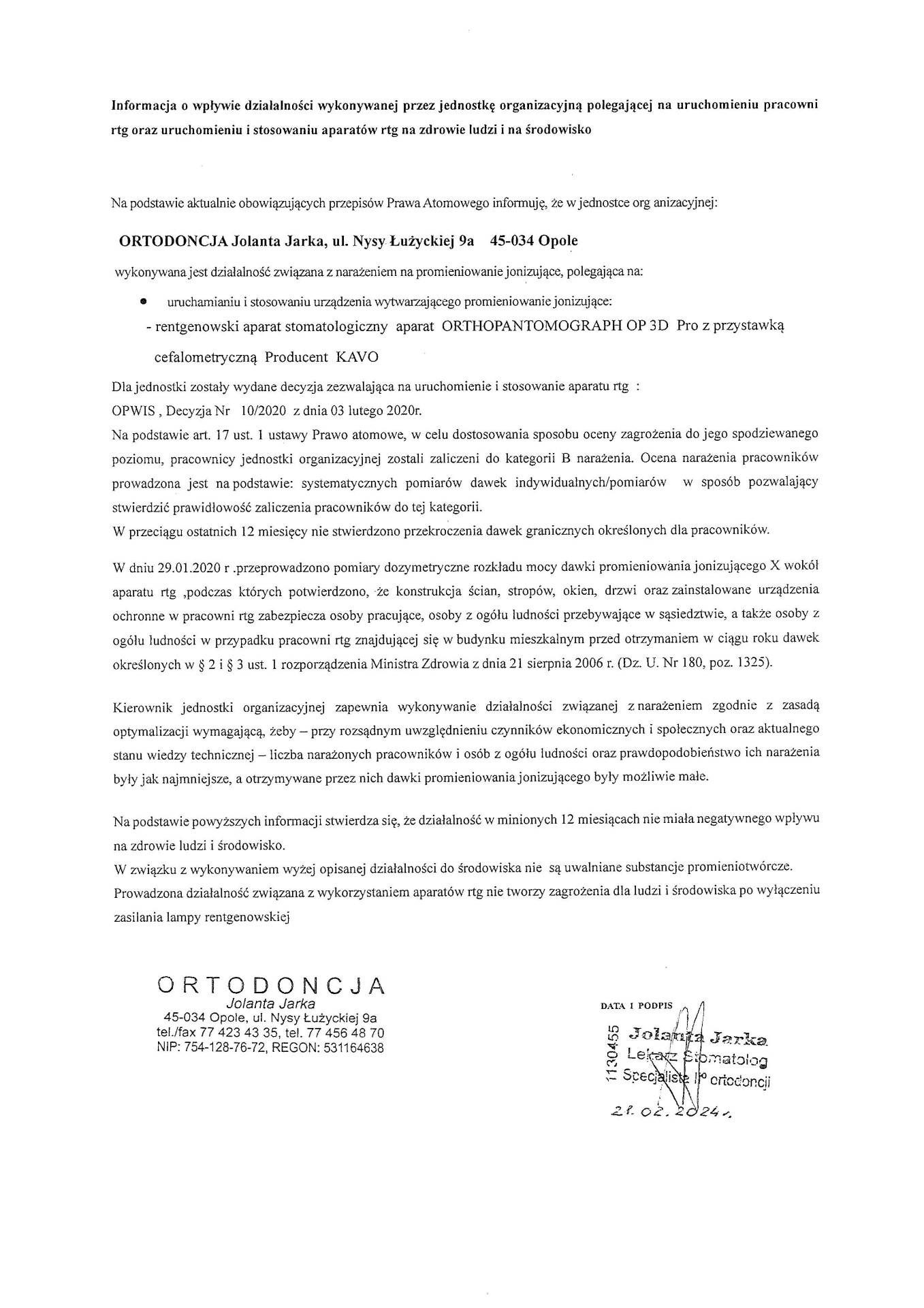
In January, we equipped our facility with a new Orthopantomograph 3D Pro computed tomography (CT) scanner from Kavo. It succeeds our previous unit, the Orthopantomograph OP 200 from the Finnish company Instrumentarium. It is worth mentioning that in 1997, we were the first orthodontic practice in the Opole region to establish our own X-ray facility, for our convenience and, naturally, that of our patients. For many years now, that Italian-made unit – then still an analog pantomographic machine with a cephalometric attachment – has been one of the exhibits at the world's largest Museum of X-ray Tubes, located at the Opole University of Technology.
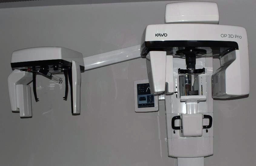
This is a versatile CT scanner featuring innovative Low Dose Technology™, and automatic dose control. This is particularly important when performing follow-up scans in children.
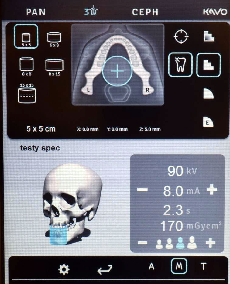
This unit produces precise 2D images thanks to its multilayer function and V-shaped X-ray beam technology. Scans can be performed in 4 image resolutions in 3D mode. It features metal artifact reduction. It also offers 5 field of view (FOV) sizes.
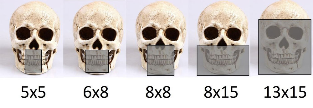
For all five fields of view, one of three resolution modes can be selected. For the 5x5 scan, an Endo mode is also additionally available.
5x5
Single implant planning
6x8
8x8
Visualization of both dental arches and portions of the maxillary sinuses:
8x15
13x15

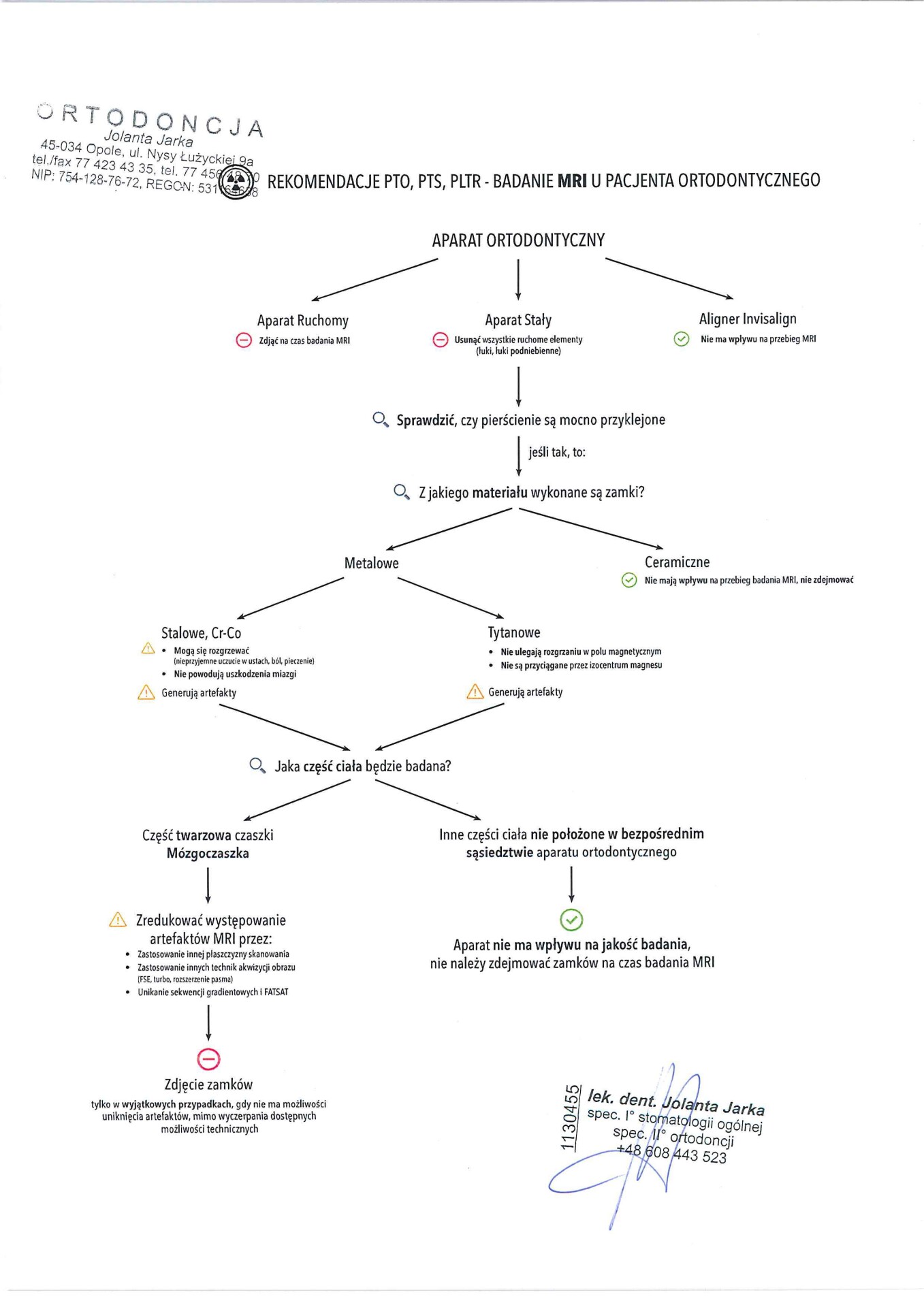
Recommendations of the Polish Orthodontic Society, Polish Dental Association, Polish Medical Radiological Society – MRI (magnetic resonance imaging) examination in an orthodontic patient
We use cookies
We use cookies to make your experience on our websites better, as well as for statistical and advertising purposes. By not blocking these files, you consent to their use and storage on your device. Please remember that you can change your browser settings to block cookies at any time. For more information, please see our privacy policy.