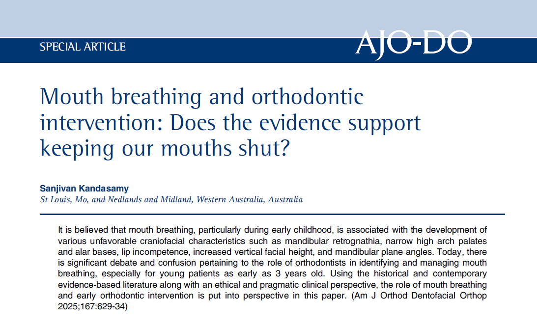
An article by Dr. Kandasamy titled 'Mouth breathing and orthodontic intervention; Does the evidence support keeping our mouths shut?' was published in AJO-DO.
https://www.ajodo.org/article/S0889-5406(25)00060-5/pdf
I read the arguments presented therein with interest; however, I do not agree with some of the points and conclusions raised. Therefore, I took the liberty of writing a response in which I present a different perspective on the discussed issues.
The issues surrounding orthodontic prophylaxis, early intervention, and early treatment have been a subject of numerous controversies for decades. The extent to which this remains poorly understood is demonstrated by the article published in Am J Orthod Dentofacial Orthop (2025;167:629-34) titled "Mouth breathing and orthodontic intervention; Does the evidence support keeping our mouths shut?" My forty years of professional experience as both a clinician and a researcher allow me to state that some of the theses presented in the article are inaccurate and inconsistent with contemporary knowledge.
It is true that mouth breathing in children can lead to significant, adverse craniofacial characteristics, such as mandibular retrognathia, a narrow and highly vaulted palate, a narrow nasal alar base, increased vertical facial height, and an increased mandibular plane angle (also known as long face syndrome).
Why? What is the main mechanism leading to these changes? Well, the prerequisite for the harmonious, mutually coordinated development of the maxilla and mandible is a sealed oral cavity. Only when the mouth is closed is it possible for the wide tongue to suction/seal to the palate (the so-called correct resting tongue position, "cobra position," or WH - vertical-horizontal). In the process of a child's growth and development from the first days, the tongue sealed to the palate stimulates its transverse growth and lowering. This, in turn, contributes to the lowering and widening of the floor of the nasal cavities and nasal passages. An open or only slightly ajar mouth causes the tongue to fall away from the palate. It rests in the mandible, and the resultant vector of the external tongue muscles is directed up and back, down, and down and forward. Thus, a muscle is missing that would counterbalance their action by pulling upwards and forwards.
The tongue must also gently rest against the dental arches to counterbalance the tension generated by the cheeks and lips from the outside of the oral cavity. Teeth erupt in a neutral space, balancing the tension of these external muscles and the tongue from the inside of the oral cavity... Instead of this "missing muscle" that maintains the tongue in the superior-anterior position, there is the mechanism of the tongue sealing to the palate, but it only works, as previously stated, when the system is sealed, i.e., the mouth is closed. When the mouth is open, the tongue's position in the oral cavity depends on which direction prevails, determined by the tension of the individual external tongue muscles. It is the tongue, a mass of strong muscles, that is primarily responsible for the direction and magnitude of mandibular growth. The mandible exhibits significantly greater morphological variability compared to the maxilla (which is more integrated with the skull bones) precisely because it is more loosely connected to the skull, being, as it were, "suspended" by the temporomandibular joints. With the mouth open, the directions and magnitude of mandibular growth and development cease to be coordinated with the growth and development of the maxilla. The maxilla and mandible grow independently, which causes skeletal disturbances.
An open mouth disrupts the entire, precisely composed (in norm), but in this dysfunction, disintegrating musculoskeletal/myofascial system. The external muscles of the tongue connect it to the skull, mandible, and hyoid bone, and through this, to the other skeletal elements, and are seamlessly integrated into the body's entire myofascial system (tensegrity).
It is very important to understand the causes of an open mouth. Therefore, a thorough history is essential.
Possible causes include:
muscle tone disorders, currently common in small and very young children (increased or decreased tone), Duchenne muscular dystrophy, and others,
respiratory system dysfunctions, such as nasal mucosal edema, allergies, asthma, hypertrophy of the pharyngeal and/or palatine tonsils, or physical obstructions,
possible habitual mouth breathing, with no discernible cause.
Yes, we propose that an open mouth should be noticed as early as possible, even in newborns, because it is always an alarm signal, indicating the presence of some dysfunction, the cause of which must be found and eliminated. Normal craniofacial development is impossible if the function of the key system, which is the respiratory system, is disrupted. The beginning of the respiratory and digestive systems, their structure, and function are closely linked, both in terms of phylogenetic development and during ontogenetic development, starting from the embryonic period.
No, we do not recommend using functional appliances in patients with laryngological problems. A child should not be forced to breathe through the nose; they should only be enabled to do so, which means we refer patients to an otolaryngologist. The otolaryngologist is the specialist competent to treat respiratory system diseases.
If the causes are muscle tone dysfunctions, we refer patients to physiotherapists and speech-language pathologists (or neurologopedists).
I agree with the statement that not all mouth-breathing patients have a retruded mandible. This situation primarily concerns patients with muscle tone disorders, habitually open mouths, and those whose laryngological cause is at the level of the palate, or the pharyngeal tonsil (allergy, asthma, nasal mucosal edema, pharyngeal tonsil hypertrophy, habitual mouth breathing, e.g., as a residual effect of frequent previous infections). These patients open their mouths, allowing the external tongue muscles to retract and lower it, which affects the magnitude and direction of mandibular growth. However, when hypertrophy of the palatine tonsils accompanies the hypertrophy of the pharyngeal tonsil, and the pharyngeal airway becomes narrowed not only in the nasal but also in the oral part, the child must open the mouth, drop the mandible, and protrude it along with the tongue to move the base of the tongue away from the posterior pharyngeal wall, thus opening the airway somewhat. Such a child has a typical adenoidal face; mid-face hypoplasia (lack of stimulating maxillary development due to the tongue resting in the mandible), dark circles under the eyes (children are unrested, oxygen-deprived, snore at night, have sleep apnea), open mouth with a protruded mandible and the tongue visible resting inside it. In these patients, a Class III skeletal malocclusion develops.
It is therefore not true that the magnitude and direction of mandibular growth are solely genetically determined.
No one disputes that the fundamentals of growth and development are genetic, nor the purpose of candidate gene studies. However, this is not synonymous with genetic determinism.
The ontogenetic development of humans, including the craniofacial complex, primarily depends on:
Genetic factors.
Environmental factors, including nutrition, biochemical interactions, and physical conditions such as pressure, temperature, hydration, and others.
Functional factors. Resulting from the mutual interaction of structure and function, including, in particular, the force and direction of muscle action Sperber G.H., Sperber SM. Craniofacial Embryogenetics and Development. Third edition, PMPH, USA 2018 .
Melvin Moss described the relationship between structure and function in the most comprehensive and scientifically substantiated way in his Functional Matrix Theory.
Therefore, it's not form over function, but rather: form follows function. If all functions within the developing craniofacial complex proceed correctly, a harmonious face will develop, exhibiting the characteristics typical of the parents. However, if dysfunctions are present (besides the previously described respiratory or muscle tone dysfunctions, also those directly related to the tongue, e.g., a significantly short lingual frenulum where the tongue cannot reach the palate), or parafunctions, there is a high probability of non-normative development. Skeletal disturbances will arise. Their symptoms may be visible in the earliest stages of development. As early as three-year-old children, we observe features of posterior, anterior, lateral functional mandibular displacement, open bite, maxillary constriction, and crossbites. It is therefore true that a new growth pattern is established as early as 4-5 years of age. Precisely because dysfunctions and parafunctions act upon a highly plastic and susceptible system—susceptible to both harmful and corrective actions. Therefore, self-correction is even possible, provided the harmful factors are eliminated. These defects will not disappear on their own. They grow with the patient, becoming more firmly anchored in the skeleton. Later, during the mixed dentition period, dental abnormalities will be added to them (these, incidentally, are indeed more strongly influenced by gene action, similar to a short lingual frenulum).
I completely agree with the statement that maxillary expansion, when there is no crossbite or transverse malocclusion in children, solely for the purpose of treating mouth breathing or sleep-disordered breathing, is unwarranted in light of scientific evidence. We, however, possess materials documenting desirable changes in palate structure in young children after the use of Hyrax-screw appliances, naturally in clinically justified cases; the palate widens and lowers. In conjunction with the elimination of the causes of open mouth (actions by otolaryngologists, physiotherapists, and speech-language pathologists/neurologopedists), this allows the child to return to the path of normal development. Such management is significantly less costly, practically free from potential iatrogenic damage, and does not require the use of retention appliances.
[Comment JJ1]: Obviously, it is not the expansion that restores the correct path, but the laryngological treatment.
Therefore, the assertion that informing parents about the harmfulness of their child's mouth breathing is "promoting disinformation and fear" or a desire to achieve greater profits, not to mention the unethical nature of such conduct, is untrue. Quite the opposite.
Every single day of our clinical work, as well as numerous scientific studies, confirms the link between laryngological problems and malocclusions. Early, sensible intervention and all interceptive measures to prevent adverse skeletal changes in a growing child should be undertaken. An orthodontist who understands the fundamentals of growth and development, what facilitates it and what is detrimental to it, has a duty to eliminate these harmful factors. Health education and prophylaxis are inherent to the medical profession. I emphasize that this is not a lucrative activity. A lot of time must be devoted to the young patient and their caregivers to clearly and understandably explain the existing problem. If the development of a malocclusion could not be avoided, then effective treatment is, of course, the final step. However, it is precisely the orthodontic treatment of skeletal malocclusions in patients who have completed their growth that requires the use of all those necessary products, appliances, and devices, and provides the greatest financial benefits to doctors, manufacturers, and distributors. I receive the suggestion that I practice prophylaxis for profit with disbelief.
Of course, I know that the article was not directed ad personam, but as the author and co-author of a publication on the development of malocclusions and how to prevent them, a guide for other specialists, a guide for parents available free of charge in our clinic and on our website, and the first quiz in the world on this topic—also available for free on the site—I felt addressed. I can guess that the intended recipients were the proponents of widespread philosophies and ideas unsupported by scientific evidence, which are related to the distribution of techniques and appliances that increase revenue, veiled by the concept of more beneficial treatment for patients, often misusing the term "holistic." For this reason, it seems worthwhile to clarify the inaccuracies, which I am doing herewith.
It is obvious that clinicians reading AJO DFO are not stuck in the knowledge of the 1860s. We cannot cure the world of retruded mandibles and mouth-breathing individuals, and it is not because we don't know how to do it. We cannot for two fundamental reasons. The first is that we treat the consequences instead of the causes. And one of the causes is the effects of civilization. The second reason is the fact that even conveying knowledge about health-damaging actions or omissions, in order to make potential patients and their children's caregivers co-responsible for their own health, does not always receive a response and compliance. Sometimes we are helpless in the face of a lack of acceptance of the simple principle that we are largely responsible for our own health. It is easier to pay for a doctor's visit than to change bad habits. That is why orthodontists will have more and more work.
Sincerely,
Jolanta Jarka
Bibliography
We use cookies
We use cookies to make your experience on our websites better, as well as for statistical and advertising purposes. By not blocking these files, you consent to their use and storage on your device. Please remember that you can change your browser settings to block cookies at any time. For more information, please see our privacy policy.